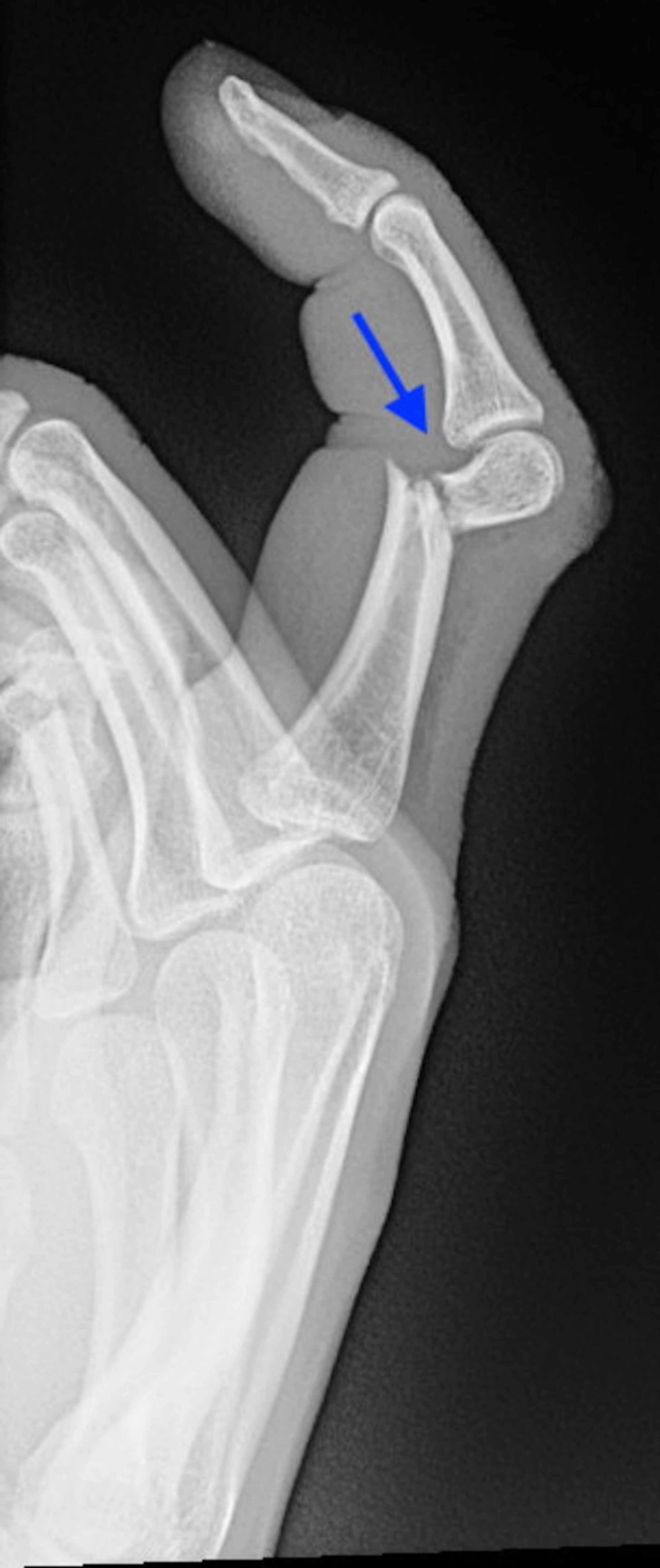

Follow-up 1 week after initial reduction with new radiographs is required to confirm the maintenance of alignment.
#Comminuted fracture hand series
Analysis of sagittal alignment on the lateral view is often difficult, particularly in plaster, and a series of oblique radiographs may be needed to confirm that correct alignment has been achieved. Post-reduction x-rays should be obtained in two planes. In the case of a stable non-displaced fracture, “buddy taping” the injured digit to an adjacent uninjured digit may be adequate. Although splinting at 90° of MCP flexion is preferable, as little as 60° of MCP flexion is likely adequate to place the collateral ligaments on maximal strain, and may be easier to achieve. Passive finger flexion is demonstrated using the tenodesis effect that occurs with passive wrist extensionĪ radial or ulnar gutter type splint with the MCP joints flexed as close to 90° as possible will hold the digits aligned while relaxing the intrinsics and preventing collateral ligament contracture. ( b) A normal finger cascade with all fingers pointing toward the thenar eminence is seen in the same patient 4 weeks after surgery.

The patient was treated with an operative reduction and percutaneous pinning. Notice the gap between the middle and ring fingers, and the deviation of the ring finger from the normal cascade. Due to a fairly normal looking appearance of the hand with the fingers extended the injury was initially treated non-operatively. ( a) This image shows a rotational malalignment of the ring finger (crossing over the small finger) from a fracture at the base of the proximal phalanx of the ring finger. Delayed treatment of these surgically indicated fractures is always more difficult, with worse functional outcomes due to stiffness, incomplete deformity correction, and post-traumatic arthritis.

Surgical treatment is indicated for any fractures of the articular surface, open fractures, fractures with significant shortening or malrotation, and fractures which fail closed reduction. These more unstable fractures require careful and frequent clinical and radiographic follow-up. Fractures with rotational or angular malaligment may be amenable to closed reduction and splinting, but these fractures are at risk for incomplete reduction and recurrent deformity. Even if splinting of one joint is needed, splints should be made small enough to allow early motion of uninjured joints.Ĭlosed non-displaced or minimally displaced fractures with acceptable alignment that are the result of a low-energy trauma usually have sufficient supporting tissues remaining intact making them stable and amenable to treatment by protected mobilization, either with local splinting of the fracture or buddy taping to adjacent fingers. For example, non-articular phalangeal fractures treated with closed reduction and splinting are mobilized after 3–4 weeks, once the fractured phalanx is less tender. Immobilization of fingers much beyond 4 weeks will lead to long-term stiffness due to extensor tendon and joint capsular scarring. Early motion prevents adhesions of the gliding soft tissues of the extensor and flexor tendon systems and prevents contracture of the joint capsules. To initiate early hand motion, fracture stability must be present either through the inherent stability of the fracture, splinting, or internal fixation. Restoration of bony anatomy is the basis for returning normal function however, an anatomic reduction is not always necessary to achieve this goal, especially if it comes at the cost of soft tissue scarring and loss of motion. The goal of treatment for any finger injury is to restore the normal function of the finger.


 0 kommentar(er)
